
How to Use Spaced Repetition for Medical Pathology
A practical guide to building high-yield pathology flashcard decks for USMLE Step 1, organized by organ system, mechanism, and histology.
Table of Contents
- Organizing Pathology Decks by Organ System and Disease Mechanism
- The Organ System Framework
- The Disease Mechanism Layer
- Card Granularity: One Fact Per Card
- Building Cards From Resources You Are Already Using
- High-Yield Pathology Content for USMLE Step 1 Preparation
- The Pareto Principle Applied to Pathology
- Building Prioritized Review Into Your Deck
- The Review Schedule That Works
- Combining Pathology Flashcards with Histology Image Review
- The Image-Integrated Card Format
- Key Histological Patterns to Master
- A Practical Histology Review Workflow
- Using LongTerMemory for Pathology Prep
- The Bottom Line
Medical pathology is one of the subjects that breaks students. Not because it is unusually abstract or deeply counterintuitive, but because of sheer volume. There are hundreds of diseases, thousands of mechanisms, genetic patterns, histological findings, and clinical correlations, and you are expected to not only know them all but to apply them rapidly under exam pressure.
The students who navigate pathology successfully almost universally have one thing in common: they have a system. Specifically, they have a spaced repetition system. Not because it is trendy, but because there is genuinely no better-evidenced approach for memorizing large volumes of factual material over an extended period.
This post is about building that system specifically for pathology, including how to structure your decks, what content to prioritize for USMLE Step 1, and how to integrate histology image review into the same workflow.
Organizing Pathology Decks by Organ System and Disease Mechanism
The single most important structural decision you will make in your pathology spaced repetition deck is how to organize it. Get this wrong and your review sessions become confusing. Get it right and the deck structure itself reinforces conceptual understanding.
The Organ System Framework
The most natural and exam-aligned organization for pathology is by organ system. This maps directly onto how Step 1 is tested and how your coursework is likely structured. Your top-level deck categories should look something like:
- Cardiovascular Pathology
- Pulmonary Pathology
- Gastrointestinal Pathology
- Hepatobiliary Pathology
- Renal and Urologic Pathology
- Hematologic and Oncologic Pathology
- Endocrine Pathology
- Reproductive and Obstetric Pathology
- Musculoskeletal and Dermatologic Pathology
- Neurologic Pathology
- Immunology and Infectious Disease
Within each organ system, organize cards around disease entities. Each disease should have a cluster of related cards covering: definition and pathogenesis, epidemiology and risk factors, clinical presentation, key laboratory or diagnostic findings, histological appearance, complications, and treatment overview.
The Disease Mechanism Layer
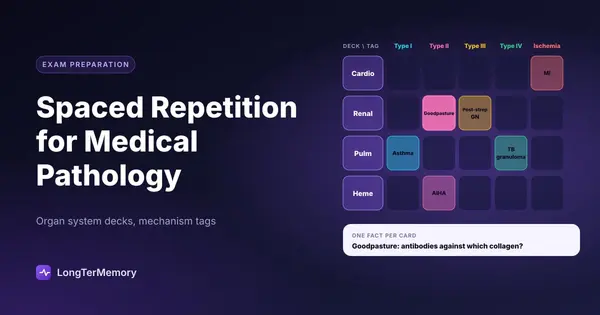
Beyond organ systems, one of the most powerful ways to add conceptual depth to your deck is to tag cards by pathophysiologic mechanism. Many diseases across different organ systems share the same underlying mechanism, and recognizing those patterns is exactly what Step 1 vignettes are designed to test.
For example, cards tagged “Type II Hypersensitivity” should span multiple organ systems: Goodpasture syndrome in the kidney, autoimmune hemolytic anemia in hematology, Graves disease in endocrinology, and so on. When you review these together, you build a mental model of the mechanism that lets you reason about novel presentations you have never explicitly studied.
| Mechanism | Example Diseases |
|---|---|
| Type I Hypersensitivity | Anaphylaxis, allergic asthma, urticaria |
| Type II Hypersensitivity | Goodpasture, autoimmune hemolytic anemia, Graves |
| Type III Hypersensitivity | Serum sickness, post-strep GN, SLE |
| Type IV Hypersensitivity | Contact dermatitis, TB granuloma, graft rejection |
| Ischemia-reperfusion injury | MI, stroke, mesenteric ischemia |
| Oncogene activation | Various cancers (KRAS, MYC, HER2) |
Card Granularity: One Fact Per Card
This is non-negotiable. A card that asks “Describe the pathogenesis, presentation, and treatment of MI” is not a flashcard. It is an essay prompt, and it will not work in a spaced repetition system because you cannot meaningfully self-grade it.
The rule: one clear question, one clear answer. Instead of a multi-part MI card, you have:
- “What is the most common cause of MI?” → Atherosclerotic plaque rupture with thrombosis
- “What enzyme rises earliest after MI?” → Troponin I/T (within 3-6 hours)
- “What is the histological finding at 1-3 days post-MI?” → Coagulative necrosis with neutrophil infiltration
- “What complication typically occurs at 3-5 days post-MI?” → Free wall rupture or papillary muscle rupture
Each of these is testable, gradeable, and retrievable as a discrete unit.
Building Cards From Resources You Are Already Using
Most medical students use First Aid for USMLE Step 1 and Pathoma as primary pathology resources. Rather than building cards from scratch, work alongside these resources. As you read a section of Pathoma, build cards in parallel. As you go through First Aid, convert high-yield facts directly into card format.
The two-step workflow: read a section, close it, try to recall the main points, then build cards for anything you could not recall cleanly. This combines initial reading with active recall and card creation in a single pass.
High-Yield Pathology Content for USMLE Step 1 Preparation
Not all pathology is equal in terms of exam frequency. Before you commit to building extensive decks, it helps to understand where the high-yield territory lies.
The Pareto Principle Applied to Pathology
A rough but useful heuristic: about 20% of pathology topics account for 80% of Step 1 pathology questions. These are the disease entities and mechanisms that appear repeatedly, often in slightly different clinical vignettes to test the same underlying knowledge.
The highest-yield categories, based on consistent NBME patterns, include:
Cardiovascular: Atherosclerosis, MI stages and complications, cardiomyopathies (dilated, hypertrophic, restrictive), endocarditis (organisms by valve and patient risk profile), cardiac tamponade, heart failure pathophysiology.
Pulmonary: Pneumonia (community vs. hospital vs. aspiration, with organisms), obstructive vs. restrictive disease patterns, lung cancers (cell type, location, paraneoplastic syndromes), TB reactivation vs. primary, pulmonary embolism.
Gastrointestinal: Crohn vs. UC (always a comparison question), liver disease progression and cirrhosis complications, hepatocellular carcinoma risk factors, celiac disease mechanism, colonic polyps and cancer.
Hematology: Anemia workup (MCV-based differential), hemoglobinopathies, coagulation factor deficiencies and their tests, lymphoma vs. leukemia classification, and hypercoagulable states.
Renal: Glomerulonephritis patterns (nephrotic vs. nephritic, causes, histology), acute tubular necrosis, renal cell carcinoma, polycystic kidney disease.
Endocrine: Thyroid disorders (hypo vs. hyper, causes, antibodies), diabetes mellitus types and complications, adrenal pathology (Cushing, Addison, pheochromocytoma), MEN syndromes.
Building Prioritized Review Into Your Deck
One practical approach: assign a priority tag (High / Medium / Low) to cards when you create them, based on how commonly the topic appears in USMLE questions. When your total review queue becomes overwhelming closer to the exam (and it will), you can filter to High priority only and get through the most important material first.
Another strategy is to use pre-made decks like Anki’s Zanki pathology deck as a starting point rather than building from scratch. The key with pre-made decks is to supplement them with your own cards for anything the deck misses or explains in a way that does not match your mental model. Never use a pre-made deck passively. Interrogate every card, edit what does not work for you, and add what you notice is missing.
The Review Schedule That Works
For Step 1 preparation, most students do best with a daily review habit rather than blocks of card review. Thirty to forty-five minutes per day of consistent review compounds dramatically over weeks. Students who skip days and then try to catch up by doing two-hour sessions are fighting the spaced repetition algorithm rather than working with it.
The algorithm is designed to show you cards just before you would naturally forget them. If you skip two days and then binge, you are reviewing some cards too early (wasted effort) and others after you have already forgotten them (playing catch-up instead of consolidating). Consistency is the whole point.
Combining Pathology Flashcards with Histology Image Review
Pathology without histology is like studying music theory without ever listening to music. The histological appearance of disease is not a separate subject from pathology. It is pathology seen at the cellular level, and Step 1 tests it in a specific and predictable way.
The Image-Integrated Card Format
The most powerful way to combine pathology and histology in your spaced repetition deck is to build image-based cards. The front of the card shows a histological image. The back gives the diagnosis, the key features visible in the image, and the relevant clinical context.
Anki supports image cards natively. You can pull histology images from Pathoma (Dr. Husain Sattar’s own drawn images are extremely useful), Robbins and Cotran Pathologic Basis of Disease, or the Ed Goljan image collection. For Step 1 specifically, focusing on images that have appeared in past USMLE exams is the most efficient strategy.
Key Histological Patterns to Master
Rather than trying to memorize every histological appearance individually, focus on pattern recognition. A small number of histological patterns recur across many diagnoses:
| Pattern | Key Features | Common Diagnoses |
|---|---|---|
| Coagulative necrosis | Cell outlines preserved, no nuclei | MI, renal infarct |
| Liquefactive necrosis | No cell outlines, pus-like | Brain abscess, bacterial pneumonia |
| Caseous necrosis | Cheese-like, granulomas | TB, fungal infection |
| Non-caseating granuloma | Epithelioid macrophages, no necrosis | Sarcoidosis, Crohn disease |
| Reed-Sternberg cells | Large cells with “owl-eye” nuclei | Hodgkin lymphoma |
| Psammoma bodies | Concentric calcifications | Meningioma, papillary thyroid Ca, serous ovarian Ca |
When you recognize a pattern rather than memorizing each disease’s histology in isolation, you can reason about unfamiliar images on the exam. “I see caseous necrosis with granulomas. That is a granulomatous inflammation pattern. In this clinical context with lung involvement and exposure history, TB is most likely.”
A Practical Histology Review Workflow
Integrate image review into your daily card sessions rather than treating it as a separate activity. A good ratio is roughly one image card for every four to five text cards. This keeps histology visible throughout your studying without it overwhelming the cognitive load of your sessions.
When you get an image card wrong, do not just accept the answer and move on. Go back to your source material and look at the image again in context. Understand what feature you missed or misidentified. Build that feature into your mental model. The next time you see a similar image, you will know exactly what to look for.
Using LongTerMemory for Pathology Prep
LongTerMemory can accelerate this process substantially. Upload your Pathoma notes, First Aid pathology sections, or annotated lecture slides, and the platform automatically generates question-answer pairs from the material. You get the benefits of active, question-based study without having to build hundreds of cards by hand.
The integrated spaced repetition scheduling means the system determines when to show you each card to maximize retention, removing the mental overhead of tracking your own review schedule. For medical students who are already managing enormous cognitive loads, outsourcing that scheduling to an algorithm is not laziness. It is smart resource allocation.
The Bottom Line
Pathology is hard because of volume, not because of complexity. Spaced repetition does not make the volume smaller. It makes your memory efficient enough to handle it.
The students who walk into Step 1 feeling confident about pathology are not the ones who read Robbins cover to cover. They are the ones who built high-yield decks, reviewed consistently for months, integrated histology into their daily practice, and let the spaced repetition algorithm do the work of scheduling.
Build the system early. Review every day. And trust the process.
The content will stick. That is the whole point of spaced repetition.